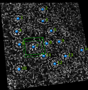

Single plane of tomogram with some virus particles marked using volume tracer.
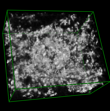
Box containing particle 7 extracted with volume dialog subregion selection. Saved to separate file for subsequent analysis.

Tom Goddard
February 15, 2008
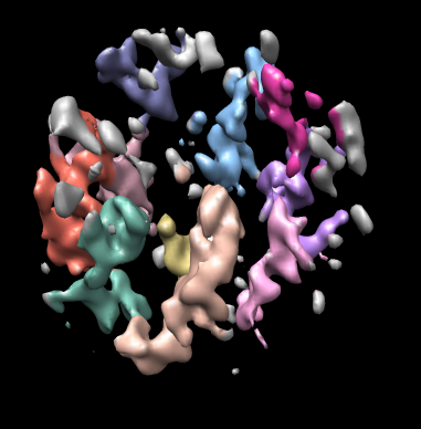
Illustration of some Chimera capabilities for segmenting electron microscope tomography. Created for Jason Lanman and Jack Johnson using their influenza virus data.
Used Chimera 1.2492, daily snapshot from February 15, 2008. Some features not available in last production release (1.2470).
The main tools used below are Volume Viewer, Volume Tracer, Color Zone, and Movie Recorder.
There are few Chimera tools for visualizing EM tomography. An extremely important one, creating masks does not exist. More capabilities for tomography are under development.
How-to style documentation is online in the Guide to Volume Data Display and in more detail in the User's Guide.

| 
|
|
Single plane of tomogram with some virus particles marked using volume tracer. |
Box containing particle 7 extracted with volume dialog subregion selection. Saved to separate file for subsequent analysis. |

|