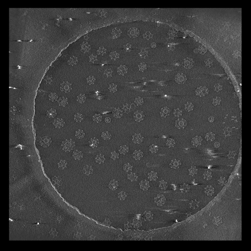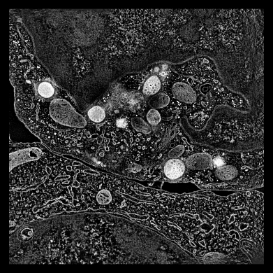
- Use volume dialog atom box to show box around center marker. 500 A pad.
- Surface display style.
- Color orange to see white specular highlights better.
|
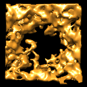
- Hide dust. Move slider to show effect.
- For unstained samples that have more dust this is especially useful.
|

- Lower threshold obscures view.
|

- Adjust box tighter (and wider) so less density blocks view.
|

- Rotate box to align with pore, allows tighter cropping.
- Rotating makes a new map interpolated from original map on rotated grid.
|

- Extract circular region of pore to examine 8-fold symmetry.
- Center marker, color zone, split map. 450 A radius.
|

- Gaussian filter pore.
- Use interactive update and adjust Gaussian width with slider. 45 A good.
|

- Find symmetry axis of pore.
- Close unneeded maps, duplicate gaussian map, rotate by hand ~180, fit map in map, "measure rotation #2 #3".
|
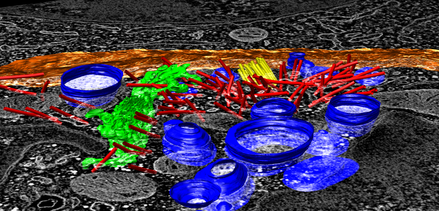
To extract density new many membrane embedded objects like 100 virus spikes
rotating a box around each is time consuming. Better to extract density
near membrane surface for the entire membrane.
To show pores in their environment make a fly-through animation.


